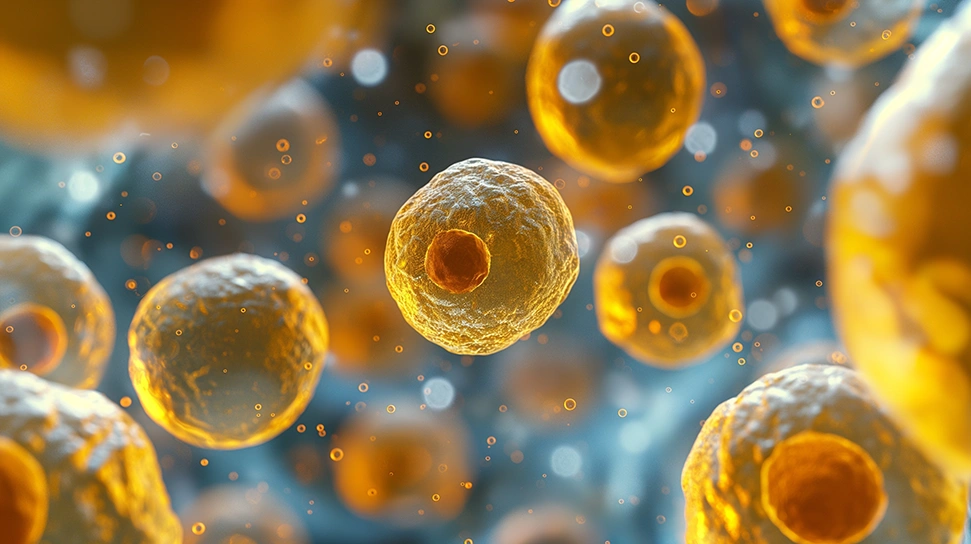
Activation is a crucial step in effective platelet rich plasma (PRP) protocols. Activation occurs after centrifugation and initiates the platelet degranulation process. Platelet degranulation releases bioactive proteins known as growth factors which increase cell mitosis, angiogenesis, chondrogenesis, and chemotaxis. Essentially, activated platelets initiate the healing cascade in damaged tissues. Some clinicians rely on local collagen type I to initiate degranulation in situ while others use calcium-based activators such as CaCl₂ or autologous thrombin. This article outlines key differences between various activation techniques especially pertaining to growth factor release rates and clot formation.
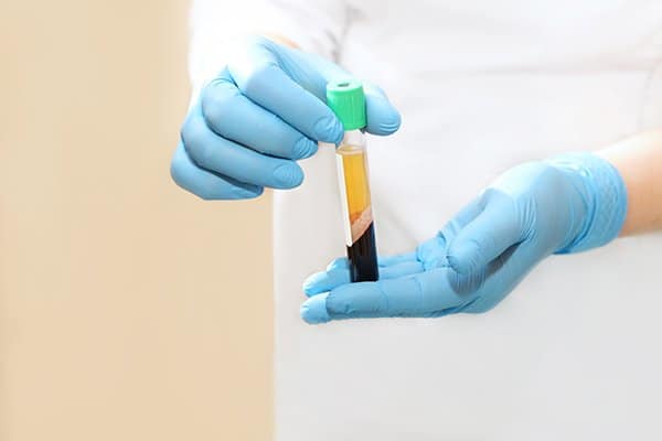
In 2016 researchers in Bologna published a comparative study which assessed the release of various growth factors after activation from one of four methods; CaCl₂, autologous thrombin, CaCl₂ with thrombin, and collagen type I.¹ Researchers collected a 150 ml blood sample from ten healthy male volunteers and prepared PRP by centrifuging the sample for 3500 rpm for 3 minutes then at 3000 rpm for 22 minutes. The PRP serum was assessed for platelet concentration and white blood cell count and then activated with 10% calcium chloride, autologous thrombin prepared from platelet poor plasma, a mixture of autologous thrombin and 10% calcium chloride, or 10% collagen type I. The activated samples were incubated at 37 degrees C (98.6 degrees F) for 15 min, 30 min, 1 hour, 2 hours, and 24 hours then centrifuged again at 2800 xg for 15 minutes. The resulting supernatants were stored at -80 degrees C until analysis.
Researchers evaluated the samples for clot formation and the release of Vascular Endothelial Growth Factor (VEGF), Transforming Growth Factor-Beta (TGF-β), and Platelet Derived Growth Factor (PDGF). Growth factor release varied at each interval depending on the activation method. Researchers found that samples activated with calcium chloride, thrombin, or CaCl₂ with thrombin formed clots starting at the 15 minute interval while samples activated with collagen type-I did not clot. Growth factor analysis showed that PRP samples activated with 10% collagen type-I released significantly less growth factors compared to other activators.
Thrombin is an enzyme that converts soluble fibrinogen into fibrin as a part of the clotting cascade. In the past, clinicians and researchers experimented with bovine thrombin as an activator, but the risk of allergic reaction is too great. Autologous thrombin can be prepared from platelet poor plasma or whole blood.² As an activator, thrombin quickly forms a dense fibrin matrix. The solidity of this fibrin matrix may inhibit cell migration.³ In the Bologna study, samples activated with thrombin released the highest levels of growth factors at 15 minutes, 30 minutes, and 1 hour, but more growth factors does not necessarily equate to a better healing response. Surrounding tissues may not have adequate time to utilize the deluge of growth factors from thrombin activation. Many growth factors have a short half life and may degrade before adequate receptors are available.

Activation from calcium chloride contributes to the formation of clots that are less dense than thrombin activated clots. The less condensed fibrin matrix may help to trap platelets and increase cell migration at the injection site. Samples activated with calcium chloride released growth factors at a significantly higher rate than collagen activated samples. Anecdotally, it seems common for medical providers in the aesthetic space to mix injectable 10% CaCl2 solution to PRP prior to facial injections. Some clinicians have suggested that the addition of a small amount of CaCl2 can help reduce the painful burning / stinging sensation that many patients experience during PRP injections. This phenomenon may be explained by the effects that CaCl2 solution has on the pH of PRP.
The combination of CaCl₂ and thrombin creates platelet rich fibrin. The calcium chloride inhibits citrate which allows the plasma to coagulate while thrombin causes fibrin to polymerize resulting in a coagulated gel. The combination results in a soft gel that can be applied in the operating room to increase post-operative wound healing. Platelets inside the soft gel will degranulate and release growth factors into surrounding tissues to increase angiogenesis, chondrogenesis and collagen secretion. Results from the Bologna study show a rapid degranulation of platelets and release of growth factors in CaCl₂/thrombin activated samples.

According to DeLong, et al. endogenous collagen activates platelets is as effectively as thrombin without forming a dense fibrin matrix.³ In his advocation of a PRP classification system, DeLong claims activation from collagen type I releases similar levels of PDFG and VEGF and greater levels of TGF-β compared to thrombin activation. The in vitro Bologna study found contrary results, but only assessed samples up to 24 hours. Collagen activated platelets did not form clots in vitro, suggesting that platelets and growth factors may migrate away from the injection site.
The researchers in Bologna utilized 10% collagen type-I to simulate in situ activation. It is common for practitioners to rely on in situ collagen to activate platelets in clinical practice. Though collagen type-I activation resulted in significantly lower growth factor release, this may be strategically advantageous depending on the type of tissue and severity of damage. Studies in mice have found excessive levels of PDGF to inhibit osteochondral regeneration.⁴ While some tissues benefit from the quick release of growth factors from exogenous activators (such as surgical openings), other cases may show poor results because growth factors can degrade before tissue receptors become available.¹
PRP activation methods dictate clot formation and rate of growth factor release. Administering effective activation protocols depends on the condition and extent of tissue damage. Some tissues benefit from the rapid release of platelets or the formation of a dense fibrin matrix, while other tissues respond to slow and gradual growth factor release. The field of orthobiologics will benefit from future studies that analyze bioactive molecule release beyond 24 hours as well as optimal concentration of activators.
Daniel Zengel, an executive with over 10 years of experience in the pharmaceutical and medical device space, is dedicated to delivering industry-leading, cost-effective products to US-based medical providers. Specializing in regenerative medicine, Daniel focuses on sales, training, and marketing support to help clinics across the country successfully implement platelet-rich plasma (PRP) therapy.